

1. INTRODUCTION
Boron Neutron Capture Therapy (BNCT) is a binary treatment modality that is based on the nuclear reaction between the stable isotope of boron (10B) and thermal neutrons, which release high-linear energy transfer (LET) ╬▒ and 7Li particles through the boron neutron capture reaction, 10B(n, ╬▒)7Li. Although most of BNCT facilities have been provided by nuclear reactors, an accelerator-based BNCT (a-BNCT) have been developed recently, because nuclear reactors regularly require long maintenance and are subject to strict regulations.
In BNCT, it is required that the irradiated dose to a patient is measured in high precision because the planning dose must be imparted accurately in tumors. In BNCT by reactors, an activation method with gold wires was popular for the purpose because it is conventional and stable. The result, however, was obtained after irradiation, not in real time. Thus the measurement with a scintillator with optical fiber (SOF) detector [1] has been proposed as a method of the real time measurement. Because the neutron beam of a-BNCT is unstable compared with that of nuclear reactors, the real time measurement is crucial to determine the planning dose. The output of the SOF detector, however, decreased with irradiation because of irradiation damage of the plastic optical fiber [2]. Hence we have developed a real-time monitor with radiation-resistant characteristics for a-BNCT.
2. DETECTOR
The conventional SOF detector consists of scintillators on the tip of two plastic optical fibers, i.e., one has a plastic scintillator with 6LiF powders on the tip for neutron detection and the other has only a plastic scintillator. Thus only neutron signals can be obtained by subtracting signals without 6LiF powders from those with 6LiF powders. The plastic scintillators are the BC490 manufactured by Bicron Ltd (Newbury, OH).
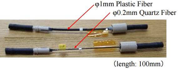
In the case of a-BNCT, the neutron fluence of 2├Ś1012 (nŌĆócm-2) is required for one treatment. In this study, for practical use of real-time monitors in a-BNCT, we set a goal that the detector can be used repeatedly, i.e, the detector has radiation tolerance for neutron fluence of 1014 (nŌĆócm-2). A quartz optical fiber is well known to have both good optical transmittance and radiation hardness compared to a plastic optical fiber. Therefore, irradiation samples were fabricated to evaluate the characteristics of radiation hardness of the quartz and plastic optical fibers. Figure 1 shows the test samples. The length of both fibers was 100 mm. The diameter of the quartz optical fiber and the plastic fiber were 0.2 mm and 1 mm, respectively. The both ends of the fibers were cut and well-polished to be connected with a standard scintillator and a measurement adapter for the X-ray irradiation.
3. IRRADIATION EXPERIMENT
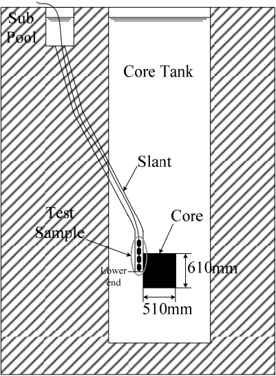
The irradiation experiment was performed at the Slant exposure tube of Kyoto University Reactor (KUR)1). Figure 2 shows the experimental setup for the irradiation in the Slant exposure tube of KUR. The size of the reactor core is approximately 51 cm├Ś51 cm├Ś61 cm (height). The reactor is a swimming pool type one, and the distance between the center of reactor core and the water surface of the core tank is approximately 6 m. The height of the lower end of the Slant exposure tube is the same as that of the center of the core. The maximum reactor power is 5 MW, while it was 1 MW in this experiment.
First of all, the distribution of the thermal neutron flux in the Slant exposure tube was measured by the activation method with gold wires. Before the measurement, we have performed calculation analysis to evaluate thermal neutron fluxes in the Slant tube with the MCNP code by modeling the KUR core and the Slant tube. Based on the evaluation, gold wires with a diameter of 0.2 mm were used in the measurement. Cd covers with a thickness of 0.5 mm were also used to measure the epi-thermal neutron distribution. The radioactivity of gold wires was measured with a high-purity Ge detector.
Figure 3 shows the measured thermal neutron flux and Cd ratio in the Slant exposure tube. The horizontal axis shows the distance between the lower end of the Slant and the measured position. The thermal neutron fluxes monotonically decreased as the distance increased. The neutron flux was in a range from 1├Ś108 to 1├Ś1012 nŌĆócm-2┬Ęsec-1. The Cd ratio was almost 20 in a distance range from 20 to 50 cm. This shows that thermal neutron was dominant in the neutron field of the Slant.
The test samples were installed and irradiated in the Slant tube. Table 1 shows the neutron flux, the neutron fluence and the irradiation positions of samples. The irradiation time was 300 seconds. The thermal neutron fluences irradiated to samples were in a range from 2.4├Ś1011 to 2.4├Ś1014 nŌĆócm-2.
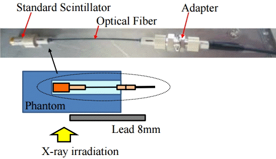
To evaluate the degradation of fibers, it is necessary to measure the transmission characteristics of optical fibers before and after the irradiation. Therefore, performance tests of samples were conducted by X-ray irradiation before and after the irradiation. As shown in Figure 4, a standard scintillator was attached to the one end of the test sample and the output signal from the scintillator was measured through the other end with a photomultiplier tube (PMT). The figure also shows the experimental setup for the X-ray irradiation. The used X-ray equipment was YXLON MG-452 (YXLON International K.K., Yokohama, Japan) and the tube voltage was 110 kV (12.5 mA). The sample fibers were set in a Polymethyl methacrylate (PMMA) acrylic phantom with a size of 30├Ś30├Ś10 cm3. Only the scintillator was irradiated with X-ray by shielding the fiber itself of the sample with lead that had a thickness of 4 mm. Thus the degradation of the fiber can be evaluated by measuring emission light from the scintillator through the fiber with a PMT. The X-ray intensity at the irradiation position was 145 mGyŌĆómin-1.
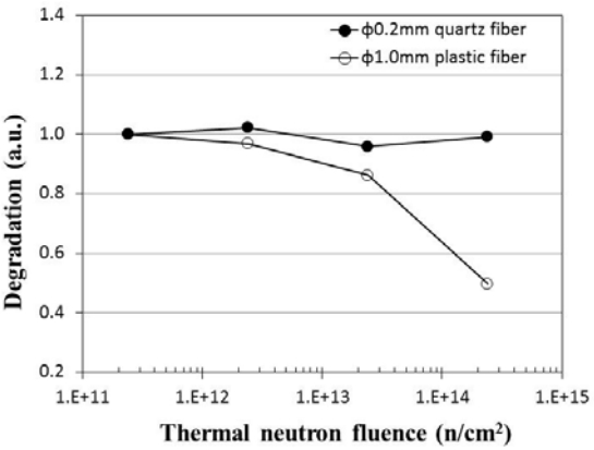
Figure 5 shows the measured results of the degradation of test samples. The degradation of the optical fibers is defined as follows:
where fafter is a count rate from the optical fiber with the plastic scintillator after the neutron irradiation experiment and fbefore is that before the irradiation. The degradation was normalized at neutron fluence of 2.4├Ś1011 nŌĆócm-2. The figure shows that the output of the plastic fiber decreased to 50% of the initial value after neutron irradiation of 2.4├Ś1014 nŌĆócm-2. On the other hand, the quartz fiber did not show any degradation in this neutron fluence. Thus it was confirmed that the quartz fiber was suitable for the detector, compared with the plastic fiber in this neutron irradiation condition.
4. IMPROVEMENT OF DETECTOR
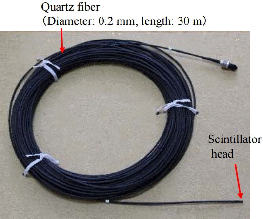
Based on the results of the irradiation experiment, new detectors consisting of quartz optical fibers were fabricated. Table 2 shows the specification of the developed SOF detector, and a photograph of the SOF detector is shown in Figure 6. The diameter of the developed quartz fiber-type SOF detector is 0.2 mm and the length is 30 m.
As for the covering material for light shielding of fiber, the materials were found to be highly activated after heavy neutron irradiation. As a result of measurement with a high-purity Ge detector, it was shown that Sb nuclide in the covering material induced the high residual activity. Therefore, in the fabrication of new SOF detectors, low activation materials that did not contain Sb nuclide were used for light shielding materials in order to avoid activation by thermal neutron irradiation.
5. PERFORMANCE EXPERIMENT
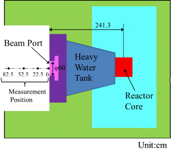
The pulse-height distribution of the developed SOF detectors was measured at Heavy Water Neutron Irradiation Facility (HWNIF) of KUR. The HWNIF can provide neutron fields with various energy spectra by changing the thickness of the heavy water. Figure 7 shows the experimental setup at the HWNIF. The distance between the center of the core and the beam port of the HWNIF is 2413 mm, the volume of heavy water tank is approximately 2 m3 and the diameter of beam port is 60 cm. To obtain high neutron intensity, we have used a beam mode, in which the fast neutrons from the reactor core were not moderated with the heavy water. The reactor power was 1MW. The thermal neutron flux was approximately 1├Ś109 nŌĆócm-2ŌĆósec-1 at the beam port, where the Cd ratio was 9.4 and the gamma dose rate was 660 mSvŌĆóh-1.
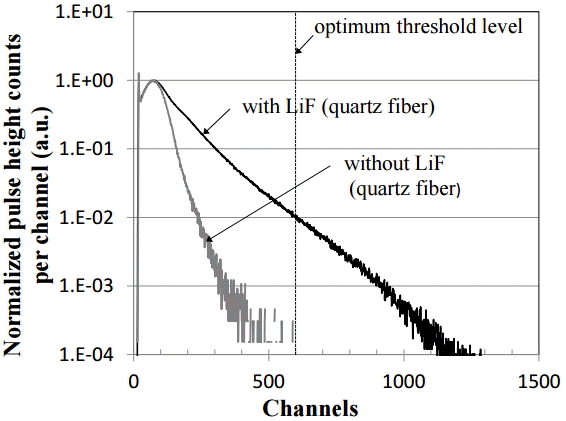
Figure 8 shows the pulse height spectrum of the signals from two fibers, i.e., one is that from a plastic scintillator with 6LiF powder and the other without the 6LiF powder, which were normalized at the peaks. As shown in Fig. 8, the signals from neutron are clearly observed over 80 channels. We can find an optimum threshold level of 600 channels to discriminate gamma-ray signals from the ratio of the pulse height spectra.
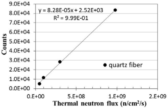
To confirm a linearity of the SOF detector, we measured the output counts by changing the intensity of thermal neutron flux, i.e., by changing the distance between the beam port and the SOF detector. The distance was changed from 0 cm to 82.5 cm and the neutron intensity was changed from 9.8├Ś108 nŌĆócm-2ŌĆósec-1 to 4.9├Ś107 nŌĆócm-2ŌĆósec-1 correspondingly. The absolute values of the thermal neutron flux were measured by the activation method with gold wires. Figure 9 shows the result of the linearity performance test. The fitting results with a least-square method showed that the SOF detector had good linearity up to about 109 (nŌĆócm-2ŌĆósec-1), where the determination coefficient was 0.999. The intensity of 109 (nŌĆócm-2ŌĆósec-1) is the same intensity of neutron fields that a-BNCT at the Ibaraki Neutron Medical Research Center (INMRC) aimed to establish [3].
6. CONCLUSIONS
We have validated the radiation hardness of quartz optical fibers for neutron fluence up to 1014 (nŌĆócm-2) and developed a new SOF detector consisting of quartz optical fibers. The discrimination characteristics of gamma-ray from neutron with the new SOF detector were well confirmed and it was also shown that the new SOF detector had good linearity in the range up to neutron intensity of 109 (nŌĆócm-2ŌĆósec-1), at which a-BNCT at INMRC aimed to establish. We are planning to conduct detailed experiments at INMRC to confirm the applicability of the new SOF detector to a-BNCT and the results will be reported in the near future. Our goal is to develop a new real-time monitor as a standard at the BNCT facility.