Introduction
Neutron spectrometers are widely used in nuclear facilities, space applications and medical applications. Information of neutron energy spectrum is required to evaluate neutron dose and also to estimate a degree of neutron activation for structural materials of facilities. Estimation of the amount of radioactive wastes activated by neutrons is quite important when decommissioning nuclear facilities. In the decommissioning works, discrimination of the wastes, in which the wastes are discriminated into radioactive or normal ones, is essential because a large amount of wastes are generated in these procedures. In order to efficiently discriminate a large amount of the wastes, the estimation of radioactive wastes based on the information of neutron flux and spectrum at interest fields is quite helpful. Although information of neutron flux and spectrum can be obtained with calculation-based estimation, measurement-based estimation is also essential for complex fields in which numerical calculations are difficult.
To obtain the neutron spectrum, Bonner sphere spectrometers (BSS) are widely used [1]. The BSS consists of several neutron detectors with different energy responses. Each detector is a thermal neutron detector such as 3He or BF3 counter surrounded with a polyethylene moderator sphere. The energy response of each detector depends on the sphere size. A large moderator sphere detector is more sensitive for fast neutrons than a small one. The BSS uses the outputs from several detectors with various size moderators to obtain a neutron spectrum. The detector outputs are the count rates obtained from each detector. The detector output vector CŌāŚ is described as
where R and ŽåŌāŚ are a detector response matrix and neutron spectral fluence, respectively. The neutron spectrum can be reconstructed by solving this equation with an unfolding technique.
In measurement procedures using the conventional BSS, the outputs from several detectors should be obtained. This means that several measurements are required at an interest measurement point. In order to reduce works for setting the detectors and measurements, a neutron spectrometer using a single detector, which has several outputs with different energy responses, is required to be developed. So far, our research group proposed and fabricated a new neutron spectrometer, which is a single moderator sphere with multiple neutron sensitive shell layers or an onion-like structure. The fabricated spectrometer has five sensitive layers. Each sensitive layers are scintillator sheets made of a LiF-ZnS mixed powder, which is sensitive only to thermal neutrons. The scintillation photons are collected with the wavelength-shifting (WLS) fiber readout, which is usually used for scintillation signal readout from large area scintillators. The scintillation sheets and WLS fibers only partially cover each shell surface because of difficulty in arranging the scintillators on the whole surface. The scintillators are uniformly and symmetrically arranged so that the detector shows no directional response dependence. The fabricated spectrometer can show appropriate outputs depending on neutron spectra and uniform directional response due to its geometric symmetry. However the spectrometer has difficulty in setting the discrimination signal level and determining the sensitivity This is because the LiF-ZnS scintillator is opaque and shows no characteristic shape, such as a peak, in a signal pulse height spectrum [2]. We, therefore, propose to replace the scintillator into a Transparent RUbber SheeT type Eu:LiCaAlF6 (TRUST Eu:LiCAF) scintillator (6Li, 95%) [3, 4]. This scintillator is flexible so that it is relatively easy to fabricate the sensitive shell layers. In this paper, we fabricate the prototype single moderator sphere neutron spectrometer with five sensitive layers using the TRUST Eu:LiCAF scintillator. We also evaluate the discrimination ability between neutrons and gamma rays. In addition, the directional response uniformity is confirmed through basic experiments using an Am-Be neutron source.
Materials and Methods
1. Details of the fabricated neutron spectrometer
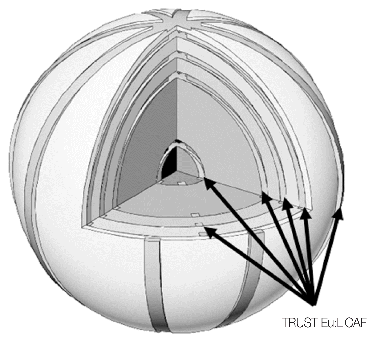
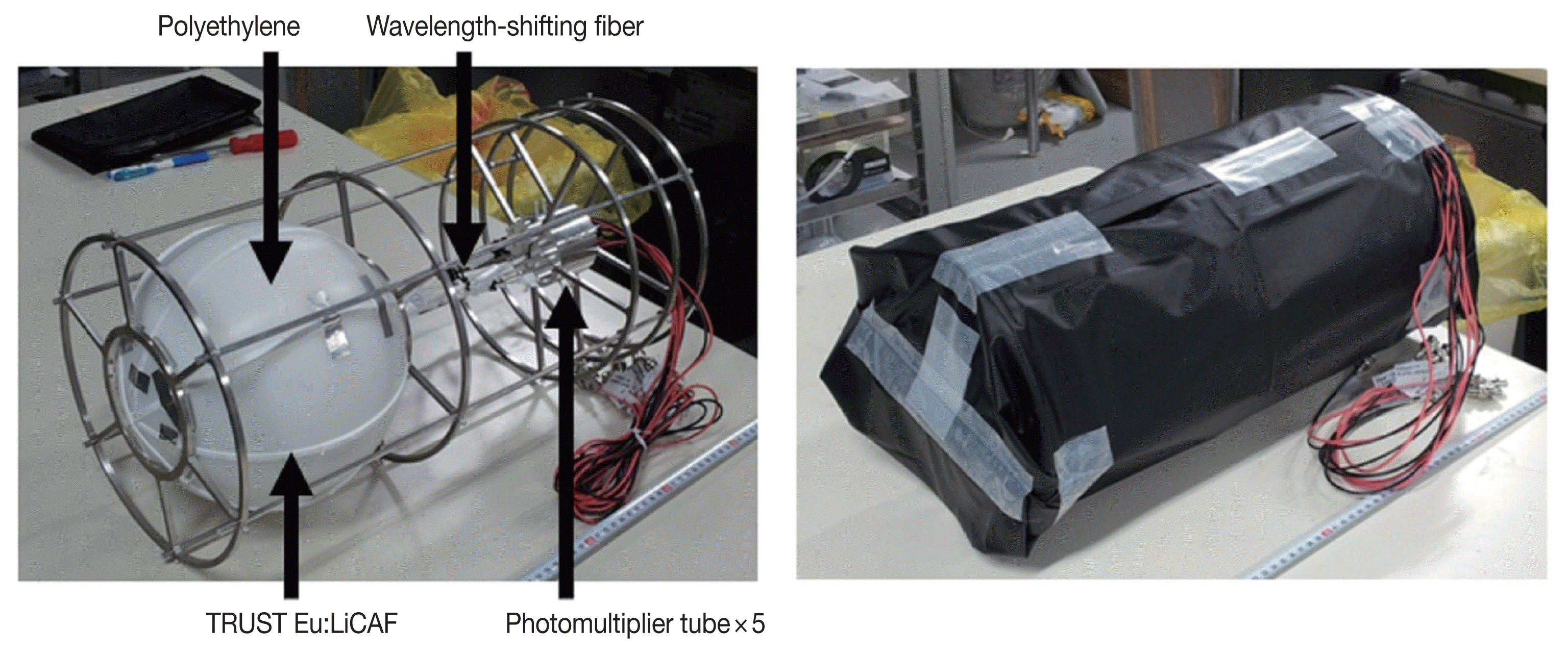
The proposed detector is a single moderator sphere with five sensitive shell layers. Figure 1 shows the schematic drawing of the proposed single sphere neutron spectrometer with multiple sensitive layers. The moderator sphere is made of polyethylene. The outer diameter of the sphere is 216 mm. In this spectrometer, four band-shaped thermal neutron detection mediums, which consist of the TRUST Eu:LiCAF scintillator and the WLS fiber readout, are looped on each shell with equal angular intervals. The Eu:LiCAF scintillator has high lithium content for neutron detection. Although the scintillation decay is not so fast and 1.6 ╬╝s decay time, it has high light yield, high transparency and no hygroscopicity [5]. However, this scintillator suffers from gamma-ray interference due to its low ╬▒/╬▓ ratio, which is the ratio of the scintillation efficiencies between alpha particles and fast electrons. In the TRUST Eu:LiCAF scintillator, a large number of small pieces of Eu:LiCAF scintillators are dispersed in a transparent resin or rubber sheet. In size controlled small scintillators, reaction products of 6Li(n,t) reactions can deposit their whole energy but fast electrons induced by gamma rays escape out with a large fraction of original energy due to difference between each range. A large fraction of fast electron energy is deposited into the rubber sheet. Since this rubber sheet is soft and flexible, the band-shaped scintillator, which can be looped on the shell surface, is relatively easy to be fabricated. The width of the scintillator band is 5 mm. The scintillation light signal can be readout with the WLS fiber accompanied with the scintillator band. The each scintillator band is wrapped with Teflon reflection sheets, If a band-shaped scintillators with uniform thickness are looped along meridians and overlapped at the pole, the response to the polar direction increases because the surface density of lithium content near the polar region is high. In order to uniform the directional response, the thickness of the scintillator band decreases near the polar region. The thickness of the thickest point scintillator is 5 mm. The both ends of WLS fibers are bundled in each layer and connected to five photomultiplier tubes (PMTs). The whole detector is covered with a black sheet for ambient light shielding. Photographs of the fabricated neutron spectrometer are shown in Figure 2. The scintillators are located at five radial distance (2.3, 7.3, 8.8, 10.3, 10.8 cm). The density of polyethylene shell sphere is 0.96 g┬ĘcmŌłÆ3.
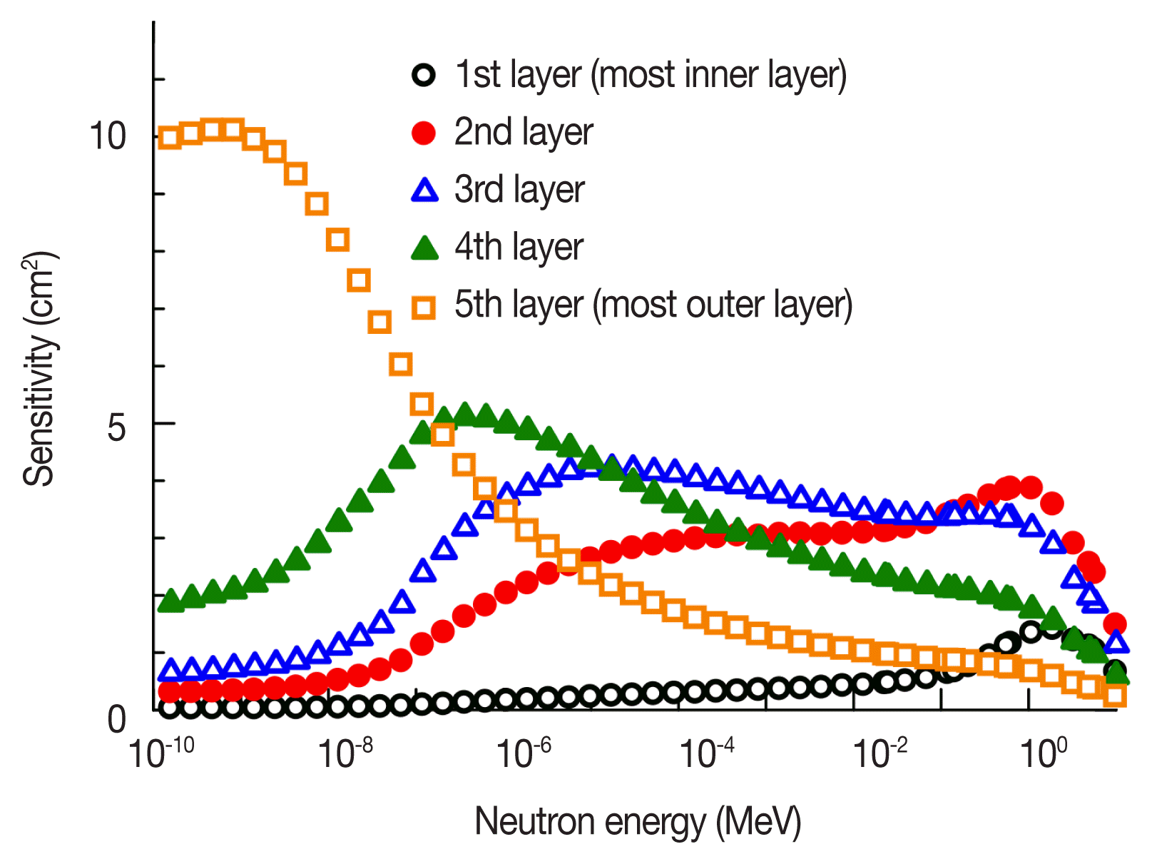
We, finally, calculated the response matrix R using Monte Carlo simulation code PHITS (Particle and Heavy Ion Transport Code System) [6]. Figure 3 shows the calculated response functions, which is the neutron energy dependences of the detector sensitivity, of each layers. The detector sensitivity was calculated as the number of 6Li(n,t) reactions per incident neutrons when the mono-energy and parallel neutron beam was uniformly irradiated over the whole detector.
2. Experimental setup
We first conducted the evaluation of neutron and gamma-ray responses of the fabricated prototype detector. For neutron irradiation experiments, a 241Am-Be neutron source of 2├Ś106 n┬ĘsecŌłÆ1 was used and the detector was placed at 1,500 mm away from the source position. For gamma-ray irradiation experiments, a 60Co gamma-ray source with 180 kBq was used and the detector was placed at 30 cm away from the source. The PMT signals are fed into signal processing circuits. Consequently, pulse height spectra are acquired with multichannel analyzers (MCAs).
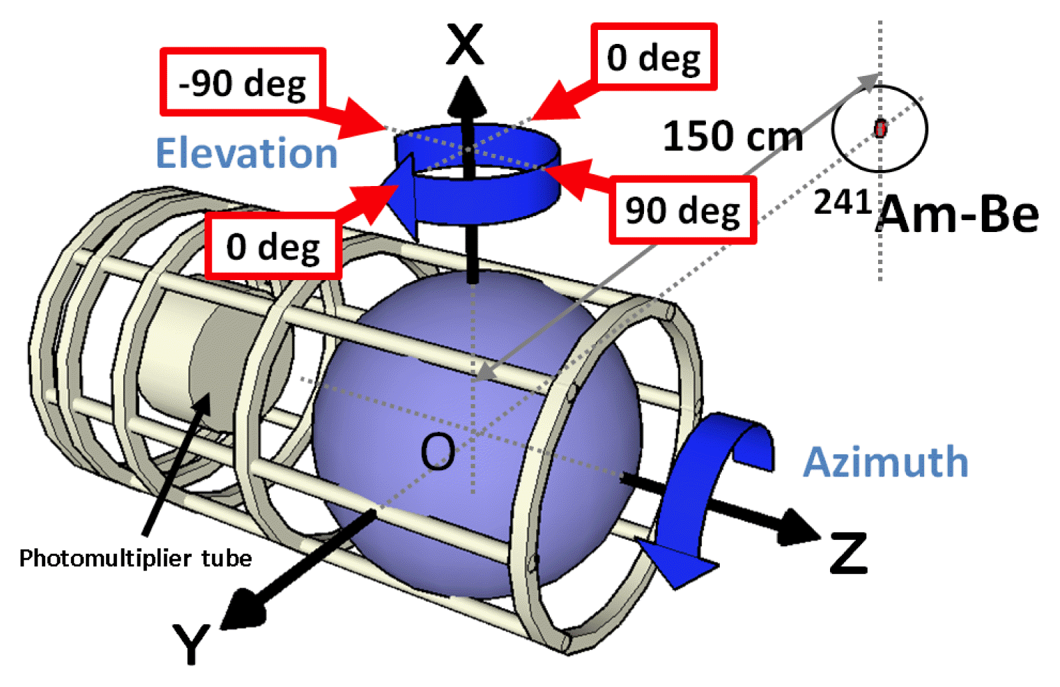
For evaluation of the directional response, we also used the 241Am-Be neutron source. In order to cancel out the influence of neutron scattering from the walls and the floor, shadow cone measurements were conducted. The polyethylene shadow cone of 250├Ś250├Ś300 mm3 was located between the neutron source and the detector to evaluate influence only of scattering neutrons. We measured the dependence of the detector sensitivity on azimuth and elevation angle of incident neutrons. The geometrical arrangement in the directional response evaluation experiments is shown in Figure 4. The elevation and azimuth angle dependence was measured by rotating the detector around the X and Z axis, respectively. The discrimination levels in the signal pulse height spectra were set for each shell layer as shown in Figure 5. The signals above the discrimination levels were recorded as neutron counts.
Results and Discussion
Figure 5 shows the pulse height spectra obtained from the neutron detection elements of each sensitive layer when irradiated with neutrons and gamma rays. Clear peaks corresponding to neutron absorption events. Therefore, we can easily determine the discrimination level. We can also confirm that the signal pulse height for gamma-ray events is preferentially suppressed. Gamma-ray events can easily be discriminated in the pulse height spectra by setting an adequate discrimination level.
Figure 6 shows the dependence of the detector sensitivity measured in experiments on elevation and azimuth angle of incident neutrons. The variation of the sensitivity is small in all directions. However, only the direction of ŌłÆ90 degrees of the elevation angle has a little bit lower sensitivity than other directions. In this direction, there are PMTs, their supporting structures and a hole in the moderator sphere for drawing out the WLS fibers. Neutrons in this direction may be interfered by the PMTs and their supporting structures. We quantitatively evaluate the sensitivity variation for elevation and azimuth angle dependence. Table 1 lists the averages and standard deviations of the detector counts of each layer. The most outer layer can strongly be affected by geometrical influence so this layer has the largest variation. For inner layers, the sensitivity modulation is milder than the most outer layer because of neutron scattering and moderation. For all detector directions, the relative standard deviations of the sensitivity in each layer are less than 12%. Excepting ŌłÆ90 degrees of the elevation angle, the standard deviation are less than 7%. The fabricated detector shows excellent directional uniformity of the neutron sensitivity.
Conclusion
We fabricated the prototype single sphere neutron spectrometer with five sensitive layers using the TRUST Eu:LiCAF scintillator and the WLS fibers. We confirmed that the fabricated detector shows a clear neutron peak and can discriminate neutron and gamma-ray events in the signal pulse height spectra. We additionally checked the directional uniformity of the detector sensitivity. The variation of the sensitivity is confirmed to be less than 12%. Excepting the direction, in which there are PMTs, their supporting structures and a hole in the moderator sphere for drawing out the WLS fibers, the variation of the detector sensitivity is less than 7%. The fabricated detector shows excellent directional uniformity of the neutron sensitivity.
As future works, we will experimentally obtain the detector response to several mono-energy neutron beams in order to check the detector response function. We should perform neutron spectrum reconstruction for a given field with a known spectrum to evaluate the performance as a neutron energy spectrometer.


