Introduction
About 900 electron linear accelerators are currently installed in hospitals in Japan, and electron acceleration energies from 6 to 15 MeV are popularly used in cancer therapy. Bremsstrahlung irradiation produced by hitting the target with accelerated electrons is the primary form of irradiation used in cancer therapies. Recently, it has become necessary to evaluate the activation of air and water to confirm that the induced activity is lower than the limit set by Japanese regulations in the new radiation protection law of radiation safety. The activation of air was induced by bremsstrahlung irradiation emitted from the target. The major activity in air is 13N (T1/2=9.96 min) from the 14N(╬│,n)13N reaction above electron energies of 10 MeV. If the bremsstrahlung irradiation hits the cooling water or water in a phantom, 15O (T1/2=122 sec) will be produced by the 16O(╬│,n)15O reaction above electron energies of 15 MeV [1].1)
For medical use, the irradiation energy is near the threshold energy of the photonuclear reaction, and the irradiation time is very short. Therefore, the induced activity of air and water does not generally cause an internal dose effect for patients and therapists because of the low induced activity and short-lived radioisotopes. We studied the activation of air and water in various accelerator facilities, which used energies between 6 and 20 MeV, in order to confirm the safety levels of activity in air in the treatment room. The concentration of radioisotopes in air and water was confirmed to be negligible below 15 MeV [2].
In this experiment, the measured activities of 13N and 15O in air and 15O in water are not consistent with the results of Monte Carlo calculations obtained using the Monte Carlo N-particle (MCNP) transport code and DCHAIN-SP code for high energy particle induced radioactivity calculation code. The measured activity was higher than calculated value. Therefore, it is important to confirm the major source of this discrepancy; for example, the accuracy of the electron energy, the Monte Carlo calculation procedure, the nuclear reaction cross-section for activation calculation, and the radioactivity measurement could be the major sources of this discrepancy. We plan to study the yields of the photonuclear reactions of light elements at the electron energies of 10 to 20 MeV at the facility for nuclear physics and to compare the results of measurement and calculation.
Materials and Methods
1. Materials
The samples were highly purified SiO2, Si3N4, and carbon disks (10 mm in diameter and 4 mm in thickness, Tokyo-Denshi Kogyo Co. Ltd., Kawasaki, Japan), which were used for monitoring the reaction yields from O, N, and C, respectively. The photon flux on each sample was monitored using Au foils (20 ╬╝m in thickness, Nilaco Co. Tokyo, Japan) by monitoring the yield of the 197Au(╬│,n)196Au reaction. The samples were sandwiched between Au monitors. The stacked samples were encapsulated in a quartz ampoule (1 mm in thickness).
2. Irradiation
An activation experiment was performed at the Research Center for Electron Photon Science, Tohoku University. The electron energy varied from 10 to 20 MeV. The electron beam current ranged from 20 to 70 ╬╝A, and the irradiation time was 10 minutes. The beam was extracted through the window of Ti foil (50 ╬╝m in thickness) and the BeO beam profile monitor (0.5 mm in thickness). The distance from the Ti window to the irradiation chamber was 19.6 cm in air. Samples and a tungsten converter were set in an irradiation chamber of stainless steel, which had cooling water circulating in the interior. Electron was converted to bremsstrahlung by hitting a tungsten plate converter (2 mm in thickness) inside the irradiation chamber, and a quartz ampoule was also set at a position 1 cm behind the converter. Irradiation setup is shown in Figure 1.
3. Measurement of radioactivity
Irradiated ampoules were transported to the radioisotope laboratory and unsealed to separate each sample and the monitors. Induced activity was measured by a portable Ge detector (GR2018, Canberra Industry Inc., Meriden, CT) coupled with MCA (Inspector 2000, Canberra Industry Inc., Meriden, CT). The ISOCS (LabSOCS) software (Canberra Industry Inc., Meriden, CT) was used for efficiency calculation. Counting was performed repeatedly. After the decay curve analysis of the full energy peak of the positron annihilation gamma ray (511 keV), the activity of 13N, 15O, and 11C was obtained at the end of irradiation.
4. Monte Carlo calculation
Monte Carlo calculation was performed by MCNP5 and Induced radioactivity calculation code, AERY (renovated DCAIN-SP2001). The Korea Atomic Energy Research Institute (KAERI) database of photonuclear cross-section data was primarily used [3]. Calculations were repeated for 1012 to 1019 electron events, which had a beam size of 3 mm in diameter. The irradiation geometry was depicted in detail. To adjust the calculated results, the measured radioactivity of the sample was corrected to the calculated position.
Results and Discussion
1. Induced activity
In case of SiO2 and Si3N4, the radioactivity induced from Si could not be observed. However, 18F activity was observed from Si3N4 irradiated from 10 to 20 MeV, and 13N activity was observed from SiO2 in the case of irradiation lower than 15 MeV. The activity of 18F seems to be caused by the impurity of fluorine, and 13N seems to be caused by contamination. Therefore, the decay analysis is important. After least squares fitting, each activity was corrected as the activity at the end of irradiation.
2. Yield of pertinent nuclear reaction
Figure 2 shows the photonuclear reaction yield curves of 13N, 15O, 11C, and 196Au from 10 to 20 MeV. All data are normalized the yield value of 196Au at 20 MeV. The measured yield curve of 196Au was in good agreement with the calculated yield. The Q-values of the 14N(╬│,n)13N, 16O(╬│,n)15O, and 12C(╬│,n)11C reactions are ŌłÆ10.6, ŌłÆ15.7, and ŌłÆ18.7 MeV, respectively. However, very low activities of 13N, 15O, and 11C were observed below the threshold energies. In this experiment, the width of the electron energy was about 5%. Therefore, the small contribution of higher energy electrons should be considered.
Figure 3 shows the relative yield to the 196Au yield. The measured yield of 13N was three to four times higher than the calculated value, and the measured yield of 15O was one order higher than the calculated value. The measured and calculated relative yield curves showed similar trends.
3. Activity in air components
In our previous work measured at the hospital [2], the activity of 13N induced in the air of the 250-L glove box under the beam exit window was 0.87 Bq┬ĘcmŌłÆ3 at 15 MeV (50 Gy, 10 minutes) irradiation. On the other hand, the calculated activity was 0.009 Bq┬ĘcmŌłÆ3. The calculation of the activity was performed with GENDLE/PD-2004 for the 14N(╬│,n)13N reaction and LA-150 for the 16O(╬│,n)15O reaction. Saeed et al. measured the activity of 13N induced in the Marinelli box filled with N2 gas at 15 MeV (45 Gy, 15 minutes) irradiation [4]. The obtained result was 0.765 Bq┬ĘcmŌłÆ3. This data is in good agreement with our data.
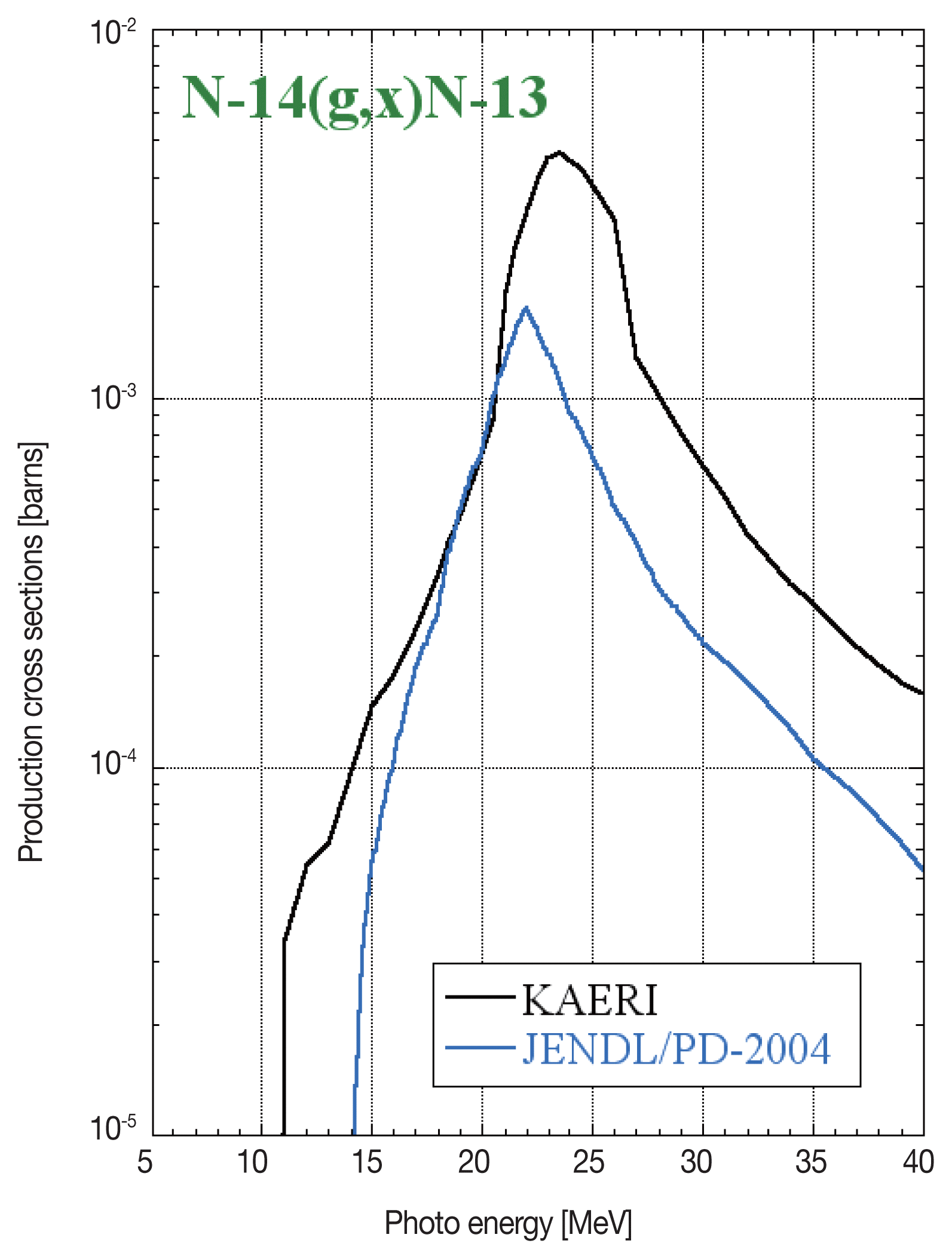
Figures 4 and 5 show the production cross-section of the 14N(╬│,n)13N and 16O(╬│,n)15O reaction, respectively. To fit the calculated data to the experimental data, the selection of cross-section data is important. In this work, we used the data set of KAERI. In this case, the discrepancy between the experiment and calculation results became small.
Conclusion
The disagreement between the experiment and calculation results might be caused by the cross-section data near the threshold energy. The experiments that obtained the photonuclear reaction cross-section for light elements were performed between 1950 and 1970. In this work using the electron accelerator for nuclear physics, it was found that the cross-section data set provided by KAERI were suited for reproducing our experimental results. Therefore, reevaluation of these results is important, especially in the threshold energy regions. Now we have a plan to recalculate by using the JENDL/PD-2015, which is newly evaluated in Japan.2) Also a spread of electron acceleration energy is unknown in case of electron accelerators for medical use. The activation of air in the accelerator room might be considered the energy spreading, because the threshold energy of photonuclear reactions of light element is near the electron acceleration energy.


